Myocardial Biopsy
What is a Myocardial Biopsy?
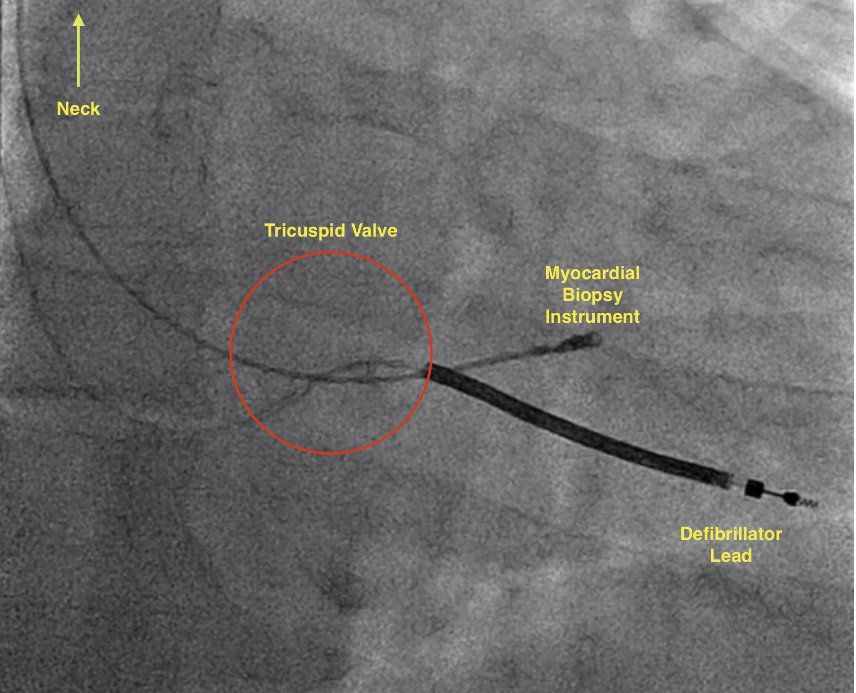
A myocardial biopsy is a medical procedure where a small sample of tissue is taken from your heart. This sample is then looked at under a microscope to check for any abnormalities or diseases. The procedure is performed using a bioptome (a pincer shaped instrument). The bioptome is inserted into a vein in your neck and gently guided to the heart.
Procedure
Under light sedation, local anaesthetic is injected into the neck near the internal jugular vein. A bioptome is introduced through the vein and guided to the heart. The ‘pincers’ of the bioptome are used to take several samples of heart muscle. The bioptome is then removed and pressure is placed on the neck to prevent bleeding. In most cases the procedure will take 10-20 minutes and you will be discharged from hospital on the same day.

Myocardial Bioptome Biopsy in a patient with a Defibrillator
Why Might You Need a Myocardial Biopsy?
- Unexplained Heart Failure: If your heart isn’t working as well as it should be, and other tests haven’t found a cause, a biopsy can sometimes reveal the diagnosis.
- Cardiac Transplant Monitoring: After a heart transplant, a biopsy can check for signs that your body might be rejecting the new heart.
- Suspected Myocarditis: If your doctor thinks you might have inflammation of the heart muscle (myocarditis), a biopsy can confirm this.
- Cardiomyopathies: These are diseases of the heart muscle. A biopsy can help identify the specific type and cause.
- To check for infections or infiltrative disorders affecting the heart.
What Are the Risks of a Myocardial Biopsy?
Like any medical procedure, there are some risks involved with a myocardial biopsy. These risks are relatively rare but include:
- Bleeding or Bruising - at the internal jugular vein
- Infection
- Pericardial effusion - bleeding around the heart that requires drainage
- Pneumothorax - a condition where air leaks into the space between the lung and the chest wall. This can happen if the catheter is inserted in the neck and accidentally punctures the lung.
- Blood Clots
- (very rare) Damage to the tricuspid heart valve